
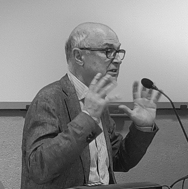
Leonardo De Chiffre, MSc, PhD, DSc
Professor Emeritus at DTU Civil and Mechanical Engineering
Applications and traceability aspects of industrial CT
Abstract
Since its introduction in the noughties, industrial CT is used for the detection of flaws and internal details in materials, and for measurements of the external as well as the internal geometry of complex parts. CT is the only technology allowing industrial quality control of workpieces with non-accessible internal features, e.g., components produced by additive manufacturing, assemblies and multi-material components. CT can be considered as a third revolutionary development in coordinate metrology, following the introduction of tactile 3D coordinate measuring machines (CMMs) in the seventies and that of optical 3D scanners in the eighties. The number of industrial applications of Computed Tomography (CT) is large and rapidly increasing. The presentation contains a survey of application examples from the manufacturing industry as well as from other industries, e.g., electrical and electronic devices, inhomogeneous materials, and from the food industry, along with applications for airport security, etc. The paramount importance of traceability of CT measurements is addressed. The paper examines procedures and artefacts to support production by establishing metrological traceability of measurements. Challenges as well as major national and international coordinated activities in the field of industrial CT are presented. Results of interlaboratory comparisons are summarized and performance in various dimensional measurement fields is illustrated.
Bio
Leonardo De Chiffre, MSc, PhD, DSc, Professor Emeritus at DTU Civil and Mechanical Engineering, Technical University of Denmark, is an internationally recognized expert in manufacturing and metrology. He has been initiator and coordinator of major framework programs and created and directed since its formation in 1992 the Centre for Geometrical Metrology (CGM) at DTU. During its activity period 1992-2016, CGM has been the foremost center of knowledge in Denmark within the field of dimensional metrology, delivering accredited calibration and testing at the highest level and operating part of the Danish National Primary Laboratory for Length under the Designated Institute DTU. Leonardo De Chiffre has also created and directed during 1992-2002 the Laboratory of Industrial and Geometrical Metrology at the University of Padova. He is Fellow of the International Academy for Production Engineering (CIRP) and Foundation Committee Member of the European Society for Precision Engineering and Nanotechnology (euspen) He is Knight of the Order of the Dannebrog and Recipient of the Euspen Lifetime Achievement Award. Leonardo De Chiffre has fostered 3 Taylor Medals from CIRP.

Dr. Antoine Klos
IMDEA Materials Institute, Madrid, Spain
Quantifying Local Information Loss in AI-Boosted X-Ray Tomography
Abstract
X-ray tomography is limited by a trade-off between scan time and spatial resolution. However, there is an increasing need for faster scans, particularly for in situ testing or high-throughput acquisitions. Such gains can be obtained by: (i) reducing exposure times, which increases the noise level; (ii) reducing the spatial sampling, which degrades the minimum resolvable feature size. In any case, it will result in information loss at the microstructure scale.
To compensate for this loss occurring during imaging, researchers proposed various post-processing strategies based on advances in deep learning for scientific imaging. In this work, particular focus is placed on two approaches that tackle different dimensions of the spatiotemporal bottleneck. On the one hand, so-called super-resolution algorithms exploit the colocalization of multi-resolution data to enhance the sharpness of low-resolution, large-field-of-view scans [1, 2]. On the other hand, state-of-the-art denoising methods enable working with previously unusable short exposure times [3].
Although both methods could help reduce scan time by several orders of magnitude, their relevance is generally quantified only in terms of grey-level similarity to a reference, using metrics such as Peak Signal-to-Noise Ratio (PSNR) or Structural Similarity Index (SSIM). Such criteria are not well-suited for materials scientists, who instead evaluate these methods based on their ability to capture metrics of interest in their field (phases, pores, roughness, etc.) or specific phenomena (coalescence, cracks, etc.).
Hence, this work will aim to go a step further by quantifying the amount of local information loss induced by both strategies. To this end, several case studies will be investigated, covering topics such as defect detection and quantification in lattice materials and microstructure characterisation of alloys. The workflows, from image acquisition to deep learning post-processing, will be detailed. Particular focus will be placed on the recovery of local features of interest, such as pores and surface roughness, enabled by these AI-boosted methods both during and after training. Finally, the relevance of each methodology will be evaluated by estimating the savings in scan time, data storage, and energy consumption.
[1] Hendriksen, A. A.; Pelt, D. M.; Palenstijn, W. J.; Coban, S. B.; Batenburg, K. J. On-the-Fly Machine
Learning for Improving Image Resolution in Tomography. Applied Sciences, 2019, 9, no. 12, 2445.
[2] Klos, A.; Salvo, L.; Lhuissier, P. Super-resolution X-ray tomography using deep learning applied to the
3D quantification of defects in lattice structures. Scientific Reports, 2025, 15, 36664.
[3] Hendriksen, A. A.; Pelt, D. M.; Batenburg, K. J. Noise2Inverse: Self-Supervised Deep Convolutional
Denoising for Tomography. IEEE Transactions on Computational Imaging, 2020, 6, 1320–35.
Bio
Antoine Klos is a postdoctoral researcher in the X-ray Characterization of Materials group at IMDEA Materials Institute in Madrid, Spain. His research focuses on the development of innovative 3D imaging methods and on the integration of artificial intelligence into scientific imaging.
He earned his PhD at the 3SR laboratory in Grenoble, France, where he investigated synchrotron X-ray microtomography in stroboscopic mode to image fast-oscillating materials at frequencies of up to several hundred hertz. Then, he held a postdoctoral position at the SIMAP laboratory in Grenoble, where he studied the application of AI to X-ray tomography, with a particular focus on defect detection and quantification in metallic lattices through super-resolution approaches.
He is currently building on this previous work to enable faster CT scans for in situ and high-throughput acquisitions, with particular attention to the quantification of information loss. His research is supported by the project METALIA – Enabling technologies for the implementation of Artificial Intelligence in the value chain of the Additive Manufacturing of new metal alloys (PLEC2023-010237, AEI, and MIG-20232050 CDTI).

Dr inż. Marcin Krawczyk, Prof. PK.
Cracow University of Technology
Challenges using XCT Systems for Coordinate Measurement
Abstract
Coordinate Measuring Systems have been used for many years to enable quick and accurate analysis of the geometric dimensions of manufactured products. Since the 1960s, we have seen an increase in their use in industry as well as in other areas of economy and culture. Recently, there has also been an increase in the use of XCT industrial tomographs to verify the geometric characteristics of industrial products. For this to be possible, it is necessary to establish traceability to the unit of length. Due to the universal nature of coordinate measurement, this is not possible through simple comparison techniques.
In the case of Coordinate Measuring Machines, the verification methods that have been developed allow the assessment of the impact of the main sources of error in these devices. Since contact measurement is much simpler than measurement using XCT systems, the use of the same procedures cannot detect the level of the main system errors.
This keynote presentation will discuss the main metrology challenges related to making accurate measurements with XCT coordinate measuring systems. The influence of various characteristic error sources associated with these systems will be discussed, together with performance verification methods.
Bio
Dr inż. Marcin Krawczyk, Prof. PK, has been dealing with issues related to coordinate measurement accuracy and methods of describing the accuracy of Coordinate Measuring Systems for over 25 years. He received his doctorate in 2005 and currently holds the position of professor at the Cracow University of Technology. Since 2010, he has been responsible for the accredited part of the Laboratory of Coordinate Metrology as an accredited calibration laboratory (currently the most accurate laboratory in Poland in the field of geometric measurements). Since 2018, he has been the delegate of the Polish Committee for Standardisation to the ISO/TC 213 Technical Committee, participating in the working groups ‘Coordinate Measuring Machines’ and ‘Uncertainty of Measurement and Decision Rules’. He has carried out numerous research projects, including European ones, and has recently been the manager of research projects related to the creation of digital twins for Coordinate Measuring Systems.

Dr Nicholas Bazin
AWE
AWE Nuclear Security Technologies Road to High Energy X-ray Computed Tomography
Abstract
High Energy X-ray Computed tomography (HEXCT) is being considered as a non-destructive testing (NDT) tool for the assessment of large, dense, components and assemblies. However, in the MeV energy regime understanding and optimising configurations and assessing/underwriting different requirements requires the development of unique Image Quality Indicators (IQI). This paper discusses our activities in designing, manufacturing, scanning, reconstructing and interpreting the respective XCT data sets from IQIs to aid in the level of confidence we can place in HEXCT imaging systems as assurance tools.
When discussing HEXCT systems we are routinely referring to 6,9 or 15 MeV linear accelerator-based sources coupled to bespoke detector panels and rotation stages housed in heavily shielded (>1m thick concrete) enclosures. Nevertheless, novel camera-based imaging, laser driven sources and developmental scintillators are also being investigated as part of our horizon scanning activities and will be briefly addressed.
To understand the art of the possible with linac-based CT systems, two, precision engineered IQIs have been manufactured and named the ‘Flowerpot’ and the ‘Minion’. Both are designed to test different aspects of high energy X-ray systems, the Flowerpot, being the most difficult to punch through, testing the higher energy range (9MeV) and the Minion bridging the gap between conventional 450keV CT systems and the low MeV range.
The Flowerpot is a continuously varying thickness split conical design incorporating numerous machined features (blind holes and groves) at varying locations (opacities) throughout the IQI. An additional central chamber comprises a tungsten cylinder and plastic core housing an inner tungsten puck with additional features machined into it.
The ‘Minion’ consists of inner and outer tungsten domes located on tungsten cylinders housed in an aluminium case. Again, similar features have been added to the surfaces of the Minion.
All features for both IQIs have been independently metrologised using CMM’s and optical metrology to provide a ‘ground truth’ against which the X-ray CT data sets can be compared. A number of different scanning criteria are also assessed for both IQI’s and will be discussed. Post scanning, reconstruction and data analysis are conducted under controlled conditions comparing measurements obtained from the XCT data and the ground truth whilst simultaneously comparing the impact of different scan criteria. Finally, these IQIs will be shipped to the US government labs where additional scanning will be carried on a quad camera-based detector system and a sub millimeter spot size 9MeV Microtron system. Once complete the IQIs will be returned to the UK where further assessment and additional features can be added if necessary.
Bio
Professor Nicholas Bazin gained his PhD at Brunel University in 1996 studying Sol-Gel Science. Following this he spent the next 15 years in the defence sector developing a number of novel materials and coatings for plasma physics experiments. It was here he was first introduced to X-ray computed tomography running a Target Fabrication Materials Development and Characterisation group where he trained up on an early X-Radia 400 mXCT instrument. Following this he was seconded to the Lawrence Livermore National Laboratories supporting collaborative UK/US research activities. On returning to the UK, he began investigating the concept of high energy radiography and computed tomography as an assurance tool. Initially looking at the concept of compact fast neutron imaging and high energy X-rays it became apparent a high energy X-ray source was the more viable option. Whilst neutrons did have unique advantages over X-rays, the maturity and ease of use of linac based x-ray systems quickly proved to be the preferred option. His program of work and his extensive team now cover multiple areas of high energy XCT development from laser driven source to scintillator development culminating in numerous national awards including the 2022 MoD Quality Award for Best Improvement Concept, 2023 MoD Chief Scientific Advisor Award and a 2024 IOM3 Defence and Security Award. Current work is focused on the design manufacture and assessment of two tungsten based IQIs comparing different systems, scanning parameters and data analysis techniques.